Use of a Smartphone for Monitoring Dermatological Lesions Compared to Clinical Photography
Dr R Asaid MBBS1, Dr G Boyce MBBS2, Dr G Padmasekara MBBS2
1Royal Melbourne Hospital, Melbourne Australia, 2Southern Health, Melbourne, Australia
Corresponding Author: raf.asaid@yahoo.com.au
Journal MTM 1:1:16-18 201
http://dx.doi.org/10.7309/jmtm.6
Background: To compare the use of a personal device such as an Apple iPhone 4 against formal clinical photography in monitoring skin lesions.
Methods: Clinical photography was used to photograph 10 skin lesions and these images were compared to photographs taken by an iPhone. These images were then reviewed by 5 different dermatologists to determine whether a discernible difference in image quality was apparent, and if sufficient detail was present to use the images taken from the iPhone in the clinical setting.
Results: All 5 dermatologists correctly identified all 10 skin lesions taken by clinical photography and those which were taken by the iPhone. Forty seven of the 50 dermatologist responses indicated both photographs provided enough detail to be clinically useful, although only 9 of the 50 responses indicated the same detail was seen in both images.
Conclusion: Although the quality of images produced using clinical photography is superior to those produced by the iPhone, pictures taken by an iPhone may provide sufficient detail for clinical assessment of skin lesions.
Introduction
Clinical photography is used in the medical profession to monitor and record various anatomical structures, wounds and lesions.1Helm TN, Wirth PB, Helm KF. Inexpensive digital photography in clinical dermatology and dermatologic surgery. Cutis 2000;65(2):103-6. While providing a useful clinical tool, costs and access can limit its use. With technological advances in portable devices such as personal cameras or smart phones with built in cameras, the ability to record and send information quickly and simple would have clear advantages over clinical photography, if image quality did not lead to a compromise in patient care.2Oakley AM, Reeves F, Bennett J, Holmes SH, Wickham H. Diagnostic value of written referral and/or images for skin lesions. J Telemed Telecare 2006;12(3):151-8. For certain lesions such as moles, clinical photography needs to be accurate and of high quality to monitor the subtle changes that occur over time. In this area, patients with multiple moles being screened for a lengthy period of time can accrue hundreds of photos, all of which require storage and come at considerable cost. Our project tests whether a picture taken on a patient’s own smartphone could be used as a cheap alternative to clinical photography, allowing patients to track and keep their own lesions.
Methods
A single subject was used to clinically document 10 skin lesions using clinical photography and an Apple iPhone 4. All lesions were first were photographed by clinical photography, with the same 10 lesions then being photographed using an iPhone.
Clinical photography uses a Nikon® D700, 105 mm macro lens, (4256×2832 pixels) and studio flash lighting. All photographs were taken following standardised views and magnifications.
Using the iPhone, we aimed to replicate similar conditions to what patients and general practitioners would be able to capture images with a well-lit room. The Apple iPhone 4 uses a 5 megapixel camera (2,592 x 1,936 pixels) and also uses a built-in high dynamic range imaging (HDR) effect, which captures three photos – one underexposed, one overexposed and one neutral – and then combines them to create an image with better dynamic range.3Apple iPhone 4 review. http://www.imaging-resource.com/PRODS/IPHONE4/IPHONE4A.HTM: Imaging Resource, 2012.
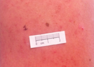
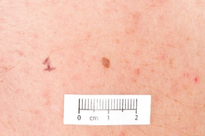
Each lesion was photographed using the Nikon D700 and the Apple iPhone 4. All images were subsequently printed to a standardised dimension of 10x15cm on photo quality paper by the same printer. An example of both images are presented in Figures 1 and 2. The pair of images were then presented to the dermatologists who were blinded to the method in which the image was taken. They were then asked the following questions:
1) Which image was the best quality?
2) Did both images have adequate detail to make a clinical decision?

Figure 1 – iPhone 4 photo of skin lesion

Figure 2 – Nikon D700 photo of skin lesion
Results
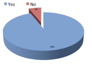
A total of 10 skin lesions were photographed with both cameras. Five dermatologists provided opinions on all 20 images. All five dermatologists found clinical photography images to be of superior quality in every skin lesion image. Forty-seven of the 50 dermatologist responses indicated both photographs provided enough detail to be clinically useful (Figure 3).

Figure 3 – “Do both photos provide adequate detail for clinical use?”
Discussion
Current literature indicates dermatologists are able to provide a more specific diagnosis with the aid of clinical photographs, in particular with regard to evaluation of inflammatory skin diseases and pigmented lesions.4Mohr MR, Indika SH, Hood AF. The utility of clinical photographs in dermatopathologic diagnosis: a survey study. Arch Dermatol 2010;146(11):1307-8. It has also been shown that dermatologists can make a correct diagnosis from an image without history,2Oakley AM, Reeves F, Bennett J, Holmes SH, Wickham H. Diagnostic value of written referral and/or images for skin lesions. J Telemed Telecare 2006;12(3):151-8. and this is true even when the lesion is not in the centre of the picture6 or the image has low resolution.5 6 This study has highlighted that although the quality of images produced using clinical photography is superior to those produced by an iPhone, this technology can still provide useful clinical information. The Apple iPhone 4, using a 5-megapixel camera can produce images that are of higher quality than standard resolution images of less than 3 megapixels, which have previously been deemed sufficient to monitor dermatological conditions.6Bittorf A, Fartasch M, Schuler G, Diepgen TL. Resolution requirements for digital images in dermatology. J Am Acad Dermatol 1997;37(2 Pt 1):195-8 A personal device such as an iPhone allows images to be kept by a patient to show to the various professionals involved in care, and images can be uploaded or sent from the device. Furthermore it offers a relatively inexpensive alternative to clinical photography, which may not be practical in certain situations such as rural and remote practice.
Further studies are needed to ascertain the ability of smartphones in monitoring lesions that are subtle and clinically challenging.
Conclusion
Smartphone cameras have progressed to an extent they are able to provide image quality, which is of sufficient detail for clinical use in dermatological practice. It must however be noted that the image quality from clinical photography is currently superior to that of an Apple iPhone 4.
References
1. Helm TN, Wirth PB, Helm KF. Inexpensive digital photography in clinical dermatology and dermatologic surgery. Cutis 2000;65(2):103-6.
2. Oakley AM, Reeves F, Bennett J, Holmes SH, Wickham H. Diagnostic value of written referral and/or images for skin lesions. J Telemed Telecare 2006;12(3):151-8. ![]()
3. Apple iPhone 4 review. http://www.imaging-resource.com/PRODS/IPHONE4/IPHONE4A.HTM: Imaging Resource, 2012.
4. Mohr MR, Indika SH, Hood AF. The utility of clinical photographs in dermatopathologic diagnosis: a survey study. Arch Dermatol 2010;146(11):1307-8. ![]()
5. Vidmar DA, Cruess D, Hsieh P, Dolecek Q, Pak H, Gwynn M, et al. The effect of decreasing digital image resolution on teledermatology diagnosis. Telemed J 1999;5(4):375-83. ![]()
6. Bittorf A, Fartasch M, Schuler G, Diepgen TL. Resolution requirements for digital images in dermatology. J Am Acad Dermatol 1997;37(2 Pt 1):195-8 ![]()

