Validation of Near Eye Tool for Refractive Assessment (NETRA) – Pilot Study
Dr Andrew Bastawrous1,2, Dr Christopher Leak2, Frederick Howard3, Mr B Vineeth Kumar1
1Wirral University Teaching Hospitals NHS Foundation Trust, UK, 2International Centre for Eye Health, Clinical Research Department, Faculty of Infectious & Tropical Diseases, London School of Hygiene and Tropical Medicine, UK, 3Independant Optometrist, UK
Corresponding Author: Andrew.bastawrous@lshtm.ac.uk
Journal MTM 1:3:6-16, 2012
DOI:10.7309/jmtm.17
Background: Uncorrected-refractive-error (URE) is the leading cause of global visionimpairment (VI); 122.5 million people are estimated VI from URE. NETRA is a $30USD clip-on application for smartphones.
Purpose: To validate the NETRA as an alternative to subjective refraction for potential use in resource-poor countries.
Methods: NETRA uses a pinhole mask attached to a smartphone displaying a spatially resolved pattern to the subject. Refractive error is estimated by the patient subjectively aligning patterns by a touchscreen interface on the smartphone. NETRA was compared to subjective refraction in 34 eyes.
Results: The mean Subjective Spherical Equivalent (SSE) was -0.65D (std 2.79, 95%CI ±0.97) Two-sided T-test showed that mean SSE is not statistically significantly different (two sided t-test; t=1.6742 p=0.1036, 95% CI±0.29) from the mean NETRA Spherical Equivalent (NSE) . Mean difference of Spherical Equivalents (NSE – SSE) was 0.24D (Std 0.84, 95%CI ±0.29). And NETRA produced a mean VA improvement of 0.44LogMAR (Std 0.52, 95%CI±0.18), or four Snellen lines.
Conclusion:In settings where access to a trained refractionist is not possible, NETRA has the potential to estimate refractive error closely enough to render an individual no longer VI from URE. NETRA is potentially a cost-effective tool in meeting the VISION2020 goals to eradicate avoidable blindness and warrants further testing in resource-poor settings.
Introduction
In 2010 the World Health Organization (WHO) announced its updated estimates of visual impairment (Snellen Acuity <6/18-3/60); globally, the number of people of all ages visually impaired is estimated to be 285 million. The principal causes being uncorrected refractive errors (URE) and cataracts, 43% and 33%, respectively. As with the Global trend in disease burden, 90% of the 122.5 million people visually impaired from URE live in low or middle income countries1Pascolini, D, Mariotti, S, Global estimates of visual impairment: 2010. Br J Ophthalmol doi:10.1136/bjophthalmol-2011-30053. Of note these figures do not include the issue of presbyopia. VISION 2020: The Right to Sight is the global initiative, of the WHO and the International Agency for the Prevention of Blindness (IAPB), to eliminate avoidable blindness by 2020. It states as one of its principle objectives, in preventing visual loss, as the need “to provide refraction and optical services that have a high success rate in terms of visual acuity and improved quality of life and are affordable, of good quality and culturally acceptable to rural, as well as urban populations”. Add to this global health inequity the conservative estimate that global productivity loss associated with URE is at $121.4 billion USD ($427.7 billion USD before adjustment for country-specific labour force participation and employment rates) and the overwhelming cost-effectiveness of refractive correction—it is possible to generate a net economic gain if the total cost of eye glasses provision were made available at less than $1000 per person2Ref: Bull World Health Organ. 2009 Jun;87(6):431-7. In most high-income countries the optometrist to population ratio is approximately 1:10,000. However, in low and middle-income countries this ratio increases dramatically to 1:600000 and much worse in many rural areas; rising to millions of people per optometrist, if one is found at all. VISION2020 recommends a minimum number for the number of trained refractionists per population (table 1)3WHO, VISION2020 Global initiative for the elimination of avoidable blindness WHO/PLB/97-61..

Table 1 – Vision2020 recommendations for number of trained refractionists per population
In most resource poor countries the number and distribution of professionally trained refractionists is not adequate to meet this unmet need4WHO Monitoring Committee for the Elimination of Avoidable Blindness VISION 2020—The Right to Sight: The Global Initiative for the Elimination of Avoidable Blindness Report of the First Meeting 2006. 17–18 January 2006. Report No WHO/PBL/06.100.. With a pair of basic prescription spectacles costing as little as $1 to produce, this lack of human resource is a major contributor to treatable visual loss due to URE in resource poor countries. Most attempts to address this unmet need have been unsuccessful, with a large reliance on volunteer organisations and overly complicated modifiable spectacles5Zhang M, Zhang R, He M, Liang W, Li X, She L, et al. Self correction of refractive error among young people in rural China: results of cross sectional investigation. BMJ 2011; 343:d4767.. Following Tudor Hart’s ‘inverses–care-law’ local optometric services tend to be private,and unevenly distributed; making them unaffordable and inaccessible to many to the urban poor and those living in rural areas. A strong current within International Health is the flow toward building health systems that are equitable, accessible and affordable to its people, and so construct an anthrogenic and sustainable health care system. The growing social acceptance and market penetration of mobile phones throughout human society has created opportunities for innovative solutions to meet this global demand to treat this unnecessary VI6Dial M for money; www.economist.com/node/9414419?story_id=9414419. Researchers at the Massachusetts Institute of Technology (MIT)7www.cameraculture.media.mit.edu/netra have developed, and are currently validating through several international collaborations (including this one), an interactive, portable, an inexpensive solution for estimating refractive errors in the human eye; the Near Eye Tool for Refractive Assessment, (NETRA). The NETRA is simply attached to a mobile phone (see figure 1) and designed to provide a rapid screening tool for refractive error. While expensive optical devices for automatic estimation of refractive error exist, NETRA simplifies the mechanism by putting the human subject in the loop. This solution is based on a high-resolution programmable display and combines inexpensive optical elements, interactive graphical user interface, and computational reconstruction. The detachable device can piggy-back on the ubiquity and penetration of mobile devices, especially in resource poor settings, so providing a possible cost effect, innovative and socially acceptable solution to VI from URE.
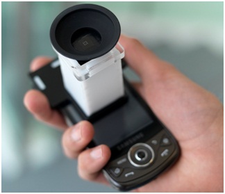
Figure 1 –NETRA Prototype attached to Samsung Behold II.
We aimed to validate the NETRA against optometric subjective refraction and calculate what proportion of individuals, classified as visually impaired (WHO) without refractive correction, would be successfully treated based on NETRA correction alone
Methods
Netra Device
The NETRA is a view dependent display to measure focusing ability of an optical system. Its basic principle exploits alignment rather than blur as an indicator of de-focus (figure 2). Trading mechanically-moving-parts for moving patterns on a digital screen, and replaces lasers, or light being focused into an eye, with a method that relies on user feedback, therefore allowing self-assessment.
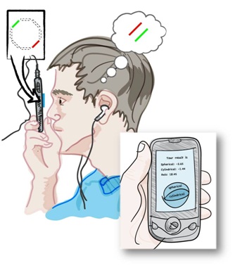
Figure 2 -Participant look at a portable touch screen display. NETRA combines inexpensive optical elements, programmable display and interactive software components to create the equivalent of a parallax barrier display that interfaces with the human eye. Using this platform, there is a new range of interactivity for measuring several parameters of the human eye, such as refractive errors, focal range, and focusing speed.
Figure 3(a) shows the basic optical setup of the NETRA system for self-evaluation of refraction. A microlens array, or a pin-hole array, is placed over a controllable high-resolution display. The viewer holds this setup in front of the eye being tested. The image formed on the viewer’s retina depends on the refractive properties of the tested eye. Using a simple interaction scheme, the user modifies the displayed pattern until the perceived image closely matches a specified result. Based on this interaction, the user’s refractive error is estimated.
Figure 3(b) shows a simplified ray diagram with two pinholes in 2 dimensions. In practice, 8 pinholes or 8 microlens (positioned on the eight neighbours of a cell centred at a 3 x 3 grid). One point is directly illuminated under each pinhole (points A and B) two parallel rays enter the eye simulating a virtual point at infinity. An eye that can focus at infinity converges these rays, which meet at a single spot P on the retina. A myopic eye however, is unable to focus at infinity, and converges these incoming rays before the retina, producing two distinct spots (PA and PB), as shown in Figure 3(c). Changing the position of point A (or B) changes the vergence of the rays produced by the pinholes. For instance, moving points A and B closer to each other on the display plane, causes the corresponding rays to diverge, progressively moving the virtual point closer to the viewer. Likewise, moving these points apart causes the associated rays to converge, moving the virtual point away from the observer. For a myopic eye, as points A and B move closer, the two imaged spots overlap on the retina at P [Figure3(c)]. The amount of shift applied to A and B allows computation of the refractive error in the viewer’s eye. The case for hyperopia is similar: as points A and B move further apart on the display plane, the resulting rays converge, creating a virtual point “beyond infinity”.8Pamplona V F. NETRA: Interactive Display for Estimating Refractive Errors and Focal Range; http://cameraculture.media.mit.edu/netra The amount of shift c required to create a virtual source at a distance d from the eye is: c = f (a/2)/ (d – t)
where t is the distance from the pinhole array to the eye, a is the spacing between the pinholes, and f is the distance between the pinhole array and the display plane. f is also the focal length of the lenslets for the micro-lens array based setup. Using a programmable LCD, the distance between the virtual scene point and the eye can be varied without any moving parts. This is equivalent to varying the power of a lens placed in front of the eye. From Equation 1, the power of a diverging lens required to fix myopia is given (in dioptres) by D = (1/d) = 1000 = (f(a/2)/c +t), where all distances are in mm. Positive values for c and D represent myopia, while negative values represent hyperopia8Pamplona V F. NETRA: Interactive Display for Estimating Refractive Errors and Focal Range; http://cameraculture.media.mit.edu/netra.
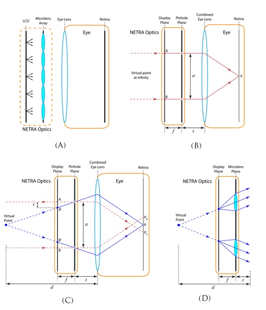
Figure 3 – NETRA optical setup: (a) a microlens array placed over a high-resolution display is held right next to the eye. For simplicity, we use a single lens to represent the combined refractive power of the cornea and the crystalline. (b) The NETRA optical system using a pinhole array. A perfect eye converges parallel rays onto a point on the retina. (c) A myopic eye converges a set of parallel rays before the retina (red arrows). By shifting points A and B to A0 and B0, respectively, the resulting rays focus on the retina at point P (blue arrows). The amount of shift required to move A to A0 allows us to compute refractive error. (d) Microlens design of the system improves the light throughput.
Performing the NETRA refraction test is a simple, quick and practicable procedure. The patient looks down the eyepiece, only being able to visualise the screen by looking directly down the eyepiece. The patient is encouraged to focus in the distance with the fellow eye. The refracting eye is presented with a pair of circular-line patterns, consisting of eight lines arranged in a circular pattern, one red the other green. Below are representations of the views for a myopic and hyperopic eye compared to emmetropia.
The patient is then instructed to slide their finger across the screen to move the circular-linear patterns until the first green and the first red lines are aligned (bottom image above). Once aligned the patient taps the screen and the phone re-presents the image with the linear pattern at a different orientation; in total eight axis powers are presented—varying by 450 each presentation. After the eight results the phone calculates the estimated NETRA prescription and presents the results on the screen as shown in figure 4.
Each Circle on the chart represents one measured value. The Left image shows 4 Readings with 0 Degrees of correction and 4 Readings around – 1.1. Negative Values mean myopia while positive means hyperopia. The Green circles shows no doubt when the patient tried to align. The Prescription is based on the best fitting curve over the data collected. Due to a very limited processor being used, the best fitting curve may be off. In This example, the subject has no myopia or hyperopia (Spherical =0), but has an astigmatism of minus 1.5 dioptres at 1530.
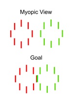
Figure 4 – Top image shows the Myopic representation; circle separate. Bottom image shows final alligment postition (Emmetropia)
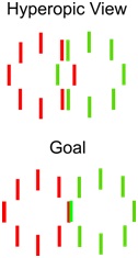
Figure 5 – Top image shows the Hyperopic representation; circle overlap. Bottom image is final position (Emmetropia).
Participants
The inclusion criteria for the study stipulated all participants must be; aged 18 years or older at the time of the examination; Phakic or pseudophakic (minimum one week post operative); able to perform subjective, objective and auto-refraction. Subjects were excluded if they had documented macular disease, or mobility/ learning difficulties rendering them incapable of using a mobile phone.
Outcomes
Test outcomes were determined by looking at three levels of examination, for correlation between the recognised Gold standard (refraction) against the NETRA. These were: (1) NETRA refraction Vs. Subjective refraction; (2) NETRA corrected visual acuity (BCNVA) Vs. Subjective (BCVA) and; (3) NETRA corrected visual acuity (BCNVA) Vs. Uncorrected Visual Acuity (UCVA).
Statistical correlation was determined as well as 95% confidence intervals and standard deviation for the mean differences.
Examination:
In total, 20 sequential patients meeting the inclusion criteria were asked to participate, three were excluded from the study. One was excluded due to bilateral age-related macular degeneration and a further two were excluded due to inability to perform the test (see discussion for more detail). In total 34 eyes of 17 patients attending a community optometrist participated. Written informed consent was given.
Due to the absence of a LogMAR acuity chart a standard backlit Snellen acuity chart was used at six meters. All subjects underwent an uncorrected Snellen visual acuity (UCVA), non-cycloplegic subjective refraction and best corrected visual acuity (BCVA) assessment by a single, senior, optometrist. Participating individuals then underwent the NETRA assessment using the NETRA and an iPod Touch (generation 4) with the assistance of the lead researcher who provided only the information supplied by MIT in two single A4 descriptive sheets. NETRA results displayed on screen were then used in a trial frame, rounding up to the nearest 0.25 dioptre, and the participants NETRA corrected visual acuity (BCNVA) was recorded.
Analysis
Data from right and left eyes in all participants were recorded and analysed (table 2). Criterions for comparison of refractive data by using differences in spherical equivalent (DSE) have been previously established.9Wesemann W, Dick B. Accuracy and accommodation capability of a handheld autorefractor. J Cataract Refract Surg 2000;26:62-70,10Schimitzek T, Wesemann W. Clinical evaluation of refraction using a handheld wavefront autorefractor in young and adult patients. J Cataract Refract Surg 2002; 28:1655-66. DSE was calculated as follows:
DSE = (SN + 0.5xCN) – (SS + 0.5xCS)
Where SN and CN represent the sphere and cylinder from NETRA refraction, and SS and CS represent the sphere and cylinder from the subjective refraction. A negative DSE indicates over minus or under plus.
Snellen visual acuity was converted to LogMAR equivalent for data analysis. Two visual acuity outcome measures were also used:
- BCVA-UCVA
- BCNVA – UCVA
Number of participants: 20 patients (40 eyes) were recruited of which 17 were examined, or 34 eyes. Validation studies of autorefractors have enrolled 100 to 200 eyes/participants and have been sufficiently powered to achieve statistical significance.11Mallen EAH, Wolffsohn JS, Gilmartin B and Tsujimura S. Clinical evaluation of the Shin-Nippon SRW-5000 autorefractor in adults. Ophthal. Physiol. Opt. 2001. Vol. 21, No. 2, pp. 101-107.,12 The limited number of participants that form the cohort of the Pilot data for this study are sufficient to inform the authors of the feasibility to pursue further funding for a larger study.
Results
Data was collected from 40 eyes, 6 eyes were excluded due to either macular degeneration (2 eyes) or inability to perform the test (4 eyes). Hyperopia was found in 15 of 34 eyes, with 3 of 34 being emmetropic and 16 of 34 being myopic. The uncorrected LogMAR equivalent of Snellen visual acuity (UCVA) and the best corrected visual acuity (BCVA) from the non-cylopegic subjective refraction assessment by the single senior optometrist, were recorded (Table 2).
|
Pt |
Age |
Eye |
UCVA |
BCVA |
|
Patient ID |
Years |
R(1) or L(2) |
Uncorrected Log Mar |
Best Subj LogMAr |
|
1 |
81 |
1 |
1.00 |
0.18 |
|
1 |
81 |
2 |
0.80 |
0.10 |
|
2 |
75 |
1 |
0.18 |
0.10 |
|
2 |
75 |
2 |
0.60 |
0.10 |
|
3 |
81 |
1 |
0.30 |
0.10 |
|
3 |
81 |
2 |
0.48 |
0.00 |
|
4 |
51 |
1 |
0.60 |
0.00 |
|
4 |
51 |
2 |
0.60 |
0.48 |
|
5 |
62 |
1 |
0.48 |
0.30 |
|
5 |
62 |
2 |
0.10 |
0.00 |
|
6 |
33 |
1 |
0.30 |
0.20 |
|
6 |
33 |
2 |
1.30 |
-0.08 |
|
7 |
42 |
1 |
1.30 |
-0.08 |
|
7 |
42 |
2 |
1.00 |
0.18 |
|
8 |
77 |
1 |
0.78 |
0.48 |
|
8 |
77 |
2 |
0.18 |
0.00 |
|
9 |
54 |
1 |
0.48 |
0.00 |
|
9 |
54 |
2 |
0.18 |
-0.08 |
|
10 |
39 |
1 |
0.18 |
0.00 |
|
10 |
39 |
2 |
0.76 |
0.00 |
|
11 |
66 |
1 |
0.60 |
0.00 |
|
11 |
66 |
2 |
0.00 |
-0.10 |
|
12 |
46 |
1 |
-0.10 |
-0.10 |
|
12 |
46 |
2 |
0.60 |
-0.10 |
|
13 |
56 |
1 |
0.48 |
-0.10 |
|
13 |
56 |
2 |
0.60 |
0.04 |
|
14 |
67 |
1 |
0.60 |
0.04 |
|
14 |
67 |
2 |
1.78 |
-0.20 |
|
15 |
30 |
1 |
1.78 |
-0.20 |
|
15 |
30 |
2 |
0.00 |
-0.15 |
|
16 |
27 |
1 |
0.18 |
-0.2 |
|
16 |
27 |
2 |
1.30 |
-0.1 |
|
17 |
23 |
1 |
1.00 |
-0.16 |
|
17 |
23 |
2 |
1.00 |
0.18 |
Table 2 – LogMAR equivalents for uncorrected and best subjective Visual acuity.
When plotted against each other the spherical equivalents for both subjective and NETRA refractions show a clear linear relationship, with a Pearson correlation coefficient, r = 0.9537 (p=0.0000), as seen in scatter plot in graph 1. The differences in spherical equivalent of the subjective optometric and NETRA tests were calculated, and the mean DSE was found to be -0.24D (Std 0.84, 95%CI ±0.29) , (Table 3)
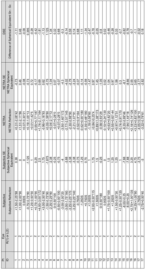
Table 3 – Subjective and NETRA prescriptions, respective spherical equivalents and DSE
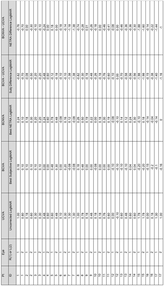
Table 4 – LogMAR equivalents for uncorrected, subjective and NETRA visual equivalents and the relative improvement produced by both refraction
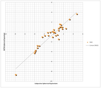
Graph 1 – A scatter plot showing the linear relationship between the NETRA-spherical-equivalent against subjective-spherical-equivalent.
Statistical analysis: A two-sided T-test showed that mean subjective spherical equivalent is not statistically significantly different from the mean of NETRA spherical equivalent (t= 1.6742 p= 0.1036, Std = 0.84, 95%CI ±0.29). These results indicate that there is no difference between the means of the two groups. (Table 4)
Best corrected visual acuities of the subjective optometric (BCVA) and NETRA (BCNVA) tests, were both recorded. A comparison of best corrected subjective (BCVA) minus uncorrected VA (UCVA), and best corrected NETRA (BCNVA) minus uncorrected VA (UCVA), was made. (Table 4)
The mean difference of subjective BCVA – UCVA was 0.58 LogMAR (Std 0.52, 95%CI ±0.18) i.e. a Snellen equivalent of 6 lines of improvement; compared to a mean difference BCNVA – UCVA of 0.44 LogMAR(Std 0.52, 95%CI±0.18) , i.e. a Snellen equivalent of 4 lines of improvement. Of the 34 eyes observed, 17 would be classed as visually impaired (Snellen VA <6/18) without correction of refractive error. 15 of these 17 (88%) eyes would no longer be classed as visually impaired based on their NETRA corrected acuity alone. In this uncorrected visually impaired group, the mean improvement in vision was 0.72 LogMar (Std 0.57, 95%CI±0.29)—equivalent to approximately 7 lines of improvement in Snellen Acuity.
A scatter plot of difference in VA from uncorrected to subjective and NETRA was also plotted. Again a clear linear relationship can be seen, with a Pearson correlation r= 0.9651 (p=0.0000), as seen in scatter plot in graph 2.
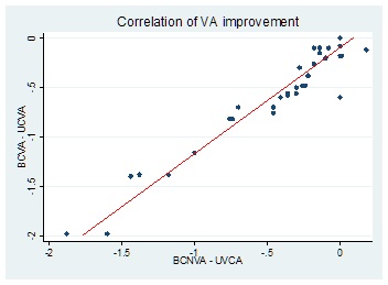
Graph 2 – A scatter plot showing the linear relationship of VA change between the Subjective and NETRA refraction.
Discussion
This pilot data shows comparable VA outcomes between NETRA and the gold standard of optometric subjective refraction. This data suggests the spherical equivalent determined by the NETRA is comparable with the spherical equivalent found by subjective refraction (mean 0.24D difference), this was more accurate in the uncorrected visually impaired eyes, with a mean difference between Subjective and NETRA refraction being 0.12D. By WHO classification of VI six subjects (35%) would have been classed as VI (person) due to uncorrected refractive error. NETRA refraction corrected their acuity to a level no longer rendering them VI.
The authors stress that the role of eye examination is not only the production of a spectacle prescription,8Pamplona V F. NETRA: Interactive Display for Estimating Refractive Errors and Focal Range; http://cameraculture.media.mit.edu/netra however to meet the growing unmet need of VI due to refractive error the validity of the NETRA as a tool for correcting URE in a setting where no refractionists are available shows promise.
The goal of this tool is not to replace the optometrists/refractionists but to be a reliable, cost-effective refractive error screening tool. Auto-refraction is typically used by refractionists as a starting point as opposed to defining the end point refraction. However, in settings where access to a trained refractionist is not possible the NETRA has the potential to estimate refractive error closely enough to render an individual no longer VI from URE. Current non-mass produced cost of the NETRA stands at only $30. The results in this pilot study are comparable to the accuracy demonstrated of marketed autorefractors.10Schimitzek T, Wesemann W. Clinical evaluation of refraction using a handheld wavefront autorefractor in young and adult patients. J Cataract Refract Surg 2002; 28:1655-66.,11Mallen EAH, Wolffsohn JS, Gilmartin B and Tsujimura S. Clinical evaluation of the Shin-Nippon SRW-5000 autorefractor in adults. Ophthal. Physiol. Opt. 2001. Vol. 21, No. 2, pp. 101-107.,12Wood MG, Mazow ML, Prager TC. Accuracy of the Nidek ARK-900 objective refractor in comparison with retinoscopy in children ages 3 to 18 years. Am J Ophthalmol. 1998 Jul; 126(1):100-8.
A Potential advantage of NETRA being connected to a mobile communication device is that it allows refraction results to be instantly displayed on the phones’ screen (Figure 6). The capacity exists to SMS the refraction to a local optical provider who can bulk make, and deliver, the spectacles to a community. Or a pre-prepared “off the shelf” spectacle can be distributed at the point of refraction13Bourne RR, Dineen BP, Huq DM, Ali SM, Johnson GJ. Correction of refractive error in the adult population of Bangladesh: meeting the unmet need. Invest Ophthalmol Vis Sci. 2004 Feb; 45(2):410-7.. This low-cost, user friendly product has the potential to contribute to the strengthening of national Eye health-care systems and facilitate national capacity-building within resource poor states, and so help VISION: 2020 realise its commitment to ‘eliminate the main causes of avoidable blindness and to prevent the projected doubling of avoidable vision impairment between 1990 and 2020 ‘.
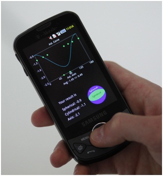
Figure 6 – Display screen for the NETRA refraction programme
Limitations of this pilot study include: we only examined 17 patients, or 34 eyes, this low number was primarily due to funding. We feel there is enough evidence now to fund expanding the number to 100 patients, or 200 eyes, in order to increase the power of the study.14Bunce, C, Correlation, Agreement, and Bland–Altman Analysis: Statistical Analysis of Method Comparison Studies. Am J Ophthalmol. 2008.09 : 2-4
NETRA had a tendency to over minus/under plus due to accommodation while performing the near task. This is largely overcome by asking the participant to open the fellow eye and fix in the distance. The observer can watch for signs on convergence which suggests the participant is accommodating and give necessary prompts. The planned NETRA prototype 2, includes a fellow eye fogging device so may overcome these issues.
Since this solution relies on subjective feedback, it cannot be used by individuals who cannot reliably perform the user-required tasks; such as very young children or someone cognitively impaired. The majority of the patients performed the self-examination after simple explanation from the assistant. However, one patient could not “see the display” even after prompting, the other Failed to understand what was being asked. Eyes that did not correct well with NETRA were mostly due to inaccurate orientation of the cylindrical axis in participants with high astigmatism (>1.50D). This is a recognised issue with autorefractors, which on the whole are only used as a starting point—very rarely will refractive correction be prescribed from an auto-refractor alone.
Further work needs to be carried out to investigate NETRA against other objective refractive instruments and its accuracy over a broader range of spectacle prescriptions. Future studies should include more examiners to generate intra-examiner reliability measurements. Future work should also assess the practicalities of this portable device in the field, and also the logistical possibility of providing the spectacles once the prescription has been ascertained.
Suggestions for full validation study
1.NETRA comparison with retinoscopy (objective refraction).
2.NETRA comparison with autorefractor (e.g. Nidek 8000).
3.Measure of accommodation.
4.In astigmatic eyes with cylinder >1.00D; measure of required steps in subjective technique to reach optimal acuity.
5.Time taken to complete test in each participant
6.Multiple examiners to assess inter observer variation
Conclusion
This device has the potential to extend an ophthalmic and public health service to people who would otherwise have very limited access, if any at all. This device can build upon the success of the well-established case finding key-informant-method15Muhit MA, Shah SP, Gilbert CE, Hartley SD, Foster A. The key informant method: a novel means of ascertaining blind children in Bangladesh. Br J Ophthalmol. 2007 Aug;91(8):995-9, to ascertain not only those who are visually impaired, or blind, but also the degree to which refractive error contributes to this. Uncorrected refractive error is the major contributor to VI globally, and places both financial and personal burden on societies that are overwhelmingly resource poor. Addressing the treatable causes of VI is a major focus (no pun intended) of the WHO and VISION2020 programme. The NETRA system provides a simple, practicable and economically valuable tool in addressing the unmet need of this major contributor to treatable global VI.
References
1. Pascolini, D, Mariotti, S, Global estimates of visual impairment: 2010. Br J Ophthalmol doi:10.1136/bjophthalmol-2011-30053 ![]()
2. Ref: Bull World Health Organ. 2009 Jun;87(6):431-7
3. WHO, VISION2020 Global initiative for the elimination of avoidable blindness WHO/PLB/97-61.
4. WHO Monitoring Committee for the Elimination of Avoidable Blindness VISION 2020—The Right to Sight: The Global Initiative for the Elimination of Avoidable Blindness Report of the First Meeting 2006. 17–18 January 2006. Report No WHO/PBL/06.100.
5. Zhang M, Zhang R, He M, Liang W, Li X, She L, et al. Self correction of refractive error among young people in rural China: results of cross sectional investigation. BMJ 2011; 343:d4767. ![]()
6. Dial M for money; www.economist.com/node/9414419?story_id=9414419
7. www.cameraculture.media.mit.edu/netra
8. Pamplona V F. NETRA: Interactive Display for Estimating Refractive Errors and Focal Range; http://cameraculture.media.mit.edu/netra
9. Wesemann W, Dick B. Accuracy and accommodation capability of a handheld autorefractor. J Cataract Refract Surg 2000;26:62-70 ![]()
10. Schimitzek T, Wesemann W. Clinical evaluation of refraction using a handheld wavefront autorefractor in young and adult patients. J Cataract Refract Surg 2002; 28:1655-66. ![]()
11. Mallen EAH, Wolffsohn JS, Gilmartin B and Tsujimura S. Clinical evaluation of the Shin-Nippon SRW-5000 autorefractor in adults. Ophthal. Physiol. Opt. 2001. Vol. 21, No. 2, pp. 101-107. ![]()
12. Wood MG, Mazow ML, Prager TC. Accuracy of the Nidek ARK-900 objective refractor in comparison with retinoscopy in children ages 3 to 18 years. Am J Ophthalmol. 1998 Jul; 126(1):100-8. ![]()
13. Bourne RR, Dineen BP, Huq DM, Ali SM, Johnson GJ. Correction of refractive error in the adult population of Bangladesh: meeting the unmet need. Invest Ophthalmol Vis Sci. 2004 Feb; 45(2):410-7. ![]()
14. Bunce, C, Correlation, Agreement, and Bland–Altman Analysis: Statistical Analysis of Method Comparison Studies. Am J Ophthalmol. 2008.09 : 2-4
15. Muhit MA, Shah SP, Gilbert CE, Hartley SD, Foster A. The key informant method: a novel means of ascertaining blind children in Bangladesh. Br J Ophthalmol. 2007 Aug;91(8):995-9 ![]()

